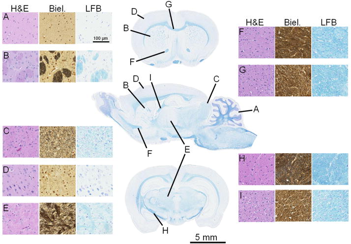
Figure 1.
A single sagittal section and two coronal sections of the normal rat brain stained with Luxol-Fast blue (center column). Anatomic structures that correspond to Table 1 are indicated (A I). For each structure, representative stains of hematoxylin and eosin (H&E), Bielchowsky’s silver impregnation (Biel.), and Luxol-Fast blue (LFB) are provided. A = cerebellar GM; B = caudate/putamen; C = superior colliculus; D = cortical GM; E = thalamus; F = anterior limb, anterior commissure; G = corpus callosum; H = internal capsule; I = ventral hippocampal commissure

