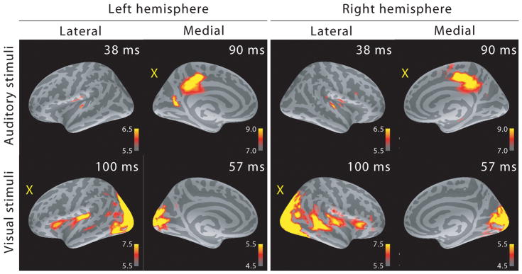
Figure 2.
MEG source analysis snapshots (dSPM F-statistics) picked at early activation latencies. Both sensory-specific and cross-sensory (marked with a yellow “X”) activations are seen (the right hemisphere calcarine cortex cross-sensory activity is not visible at this threshold). While some of the cross-sensory activations are located inside the sensory areas (as delineated in (Desikan et al., 2006)), these seem to occupy slightly different locations than the sensory-specific activations. However, the spatial resolution of MEG is somewhat limited – hence exact comparisons are discouraged. Visual checkerboard stimuli activated additional areas outside the sensory cortices, for example superior temporal sulci (STS) especially in the right hemisphere and Broca’s areas bilaterally.

