Figure 1.
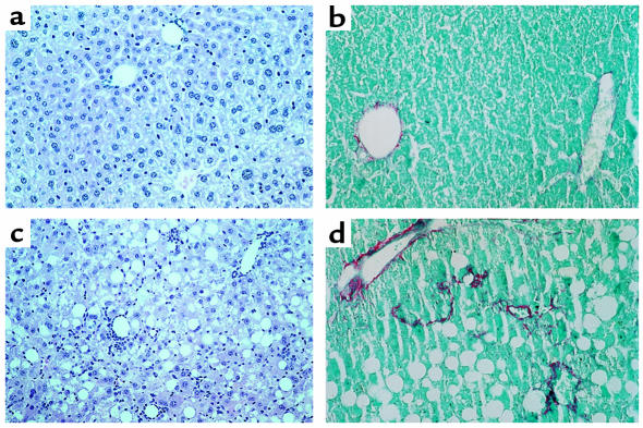
Liver sections from female C57BL6/J mice fed the MCD diet and the control diet for 10 weeks. (a) Control mouse, H&E staining (×100). (b) Control mouse, Sirius red staining (×160). (c) MCD diet–fed mouse, H&E staining (×100). In addition to the macrovesicular steatosis mainly localized in zone 2, large areas of mixed inflammatory infiltrate with lymphocyte and polymorphonuclear neutrophil necroinflammation can be seen throughout the hepatic lobule. (d) MCD diet–fed mouse, Sirius red staining (×160). The collagen fiber deposits (stained red) confirm the discrete perivenular and pericellular fibrosis.

