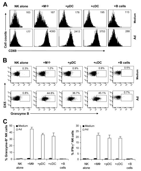
FIGURE 1.
Accessory cells are required for NK cell activation in response to adenoviral infection. Purified splenic NK cells (DX5+CD3−) were either cultured alone (NK alone), or co-cultured with peritoneal macrophages (+Mφ), pDCs (+pDCs), cDCs (+cDCs) or B cells (+B cells) in the presence (Ad) or absence (Medium) of Ad-LacZ. 18 h later, brefeldin A was added into each well and incubated for additional 4 h. (A) NK cells were analyzed for the expression of the early activation marker, CD69, by flow cytometry. The mean fluorescent intensity (MFI) of CD69 staining is indicated. Events were gated on DX5+CD3− NK cells. (B-C) NK cells were stained intracellularly for IFN-γ and granzyme B and analyzed by flow cytometry. FACS plots of granzyme B production by NK cells with the percentage of granzyme B positive cells among DX5+CD3− NK cells indicated (B). The mean percentage ± SD of granzyme B or IFN-γ positive cells among DX5+CD3− cells is indicated (n = 5 per group) (C). The data shown are representative of four independent experiments.

