
Fig. 3. MRI of hypertrophic cardiomyopathy in 46-year-old woman.
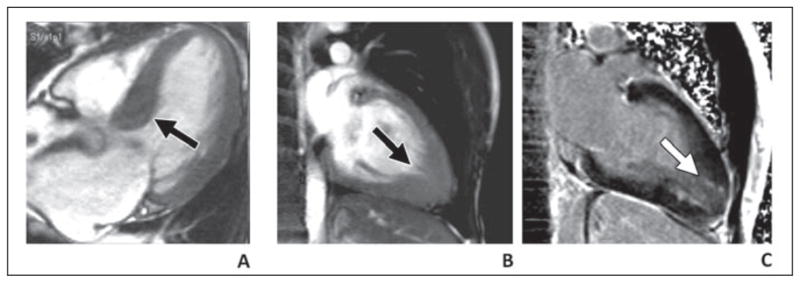
A, Septal thickening (arrow) of left ventricular outflow tract with hypertrophic obstructive cardiomyopathy. B and C, Thickening of apex (B) with myocardial delayed enhancement (C) with apical variant hypertrophic cardiomyopathy.