
Fig. 4. MRI of arrhythmogenic right ventricular dysplasia in 32-year-old man.
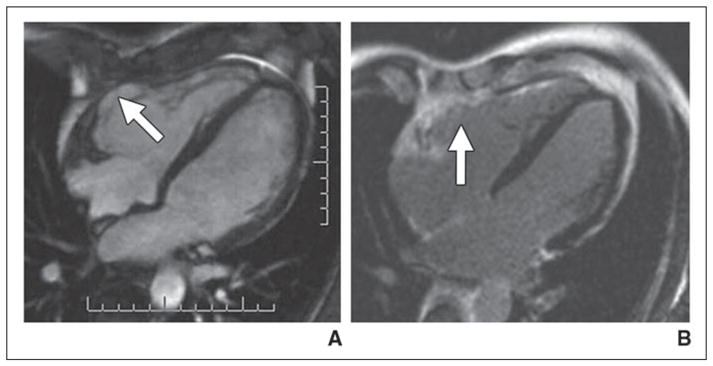
A, Axial cine steady-state free precession image shows aneurysm (arrow) involving anterior wall of right ventricle.
B, Delayed gadolinium-enhanced MRI shows enhancement (arrow) in location of right ventricular aneurysm.