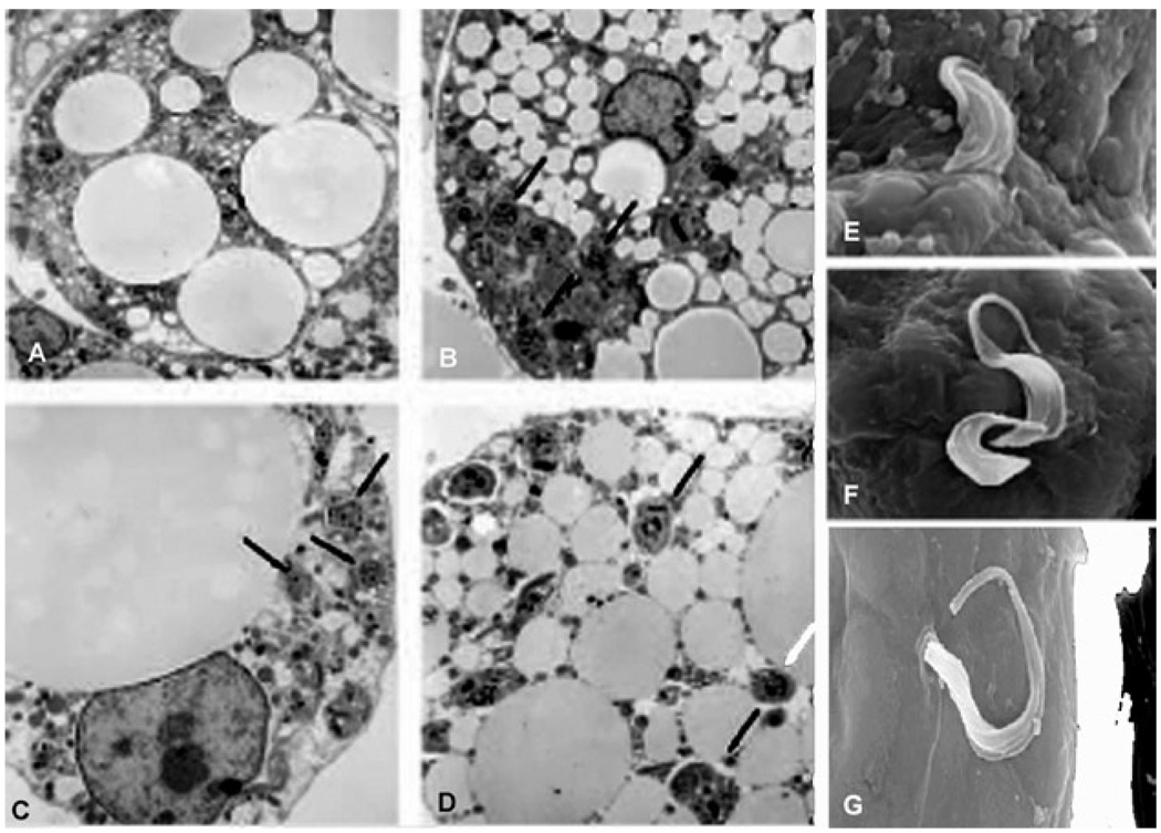
Fig. 4.
A: an uninfected cell; B–D: representative transmission electron micrographs of 3T3-L1 adipocytes 48 h post-infection. Note the close proximity of parasites to lipid droplets indicated by arrowheads; E–G: scanning electron micrographs showing invasion of adipocytes by trypomastigotes [from Combs et al. (2005) with permission of the Journal of Biological Chemistry].

