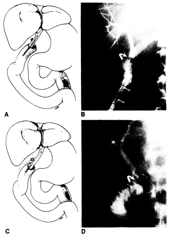
Fig. 2.
Normal choledochojejunostomy in Roux-en-Y. A, Schematic diagram shows straight internal stent across biliary anastomosis. B, PTC shows biliary anastomosis (curved arrow). Note migration of stent (arrows) into Roux limb of jejunum. C, Schematic diagram shows T-tube stent which enters donors cystic duct remnant. D, T-tube cholangiogram shows biliary anastomosis (curved arrow).

