
Fig. 4.
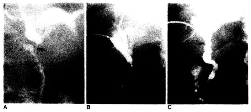
Treatment of common hepatic duct stricture by transhepatic balloon dilatation. A. T-tube cholangiogram 3 weeks after transplantation demonstrates biliary obstruction due to stricture in donor’s common hepatic duct (arrow). Note donor’s and recipient’s cystic duct remnants (arrowheads). B, Transhepatic dilatation with 8-mm balloon. C. Catheter cholangiogram 6 weeks after dilatation demonstrates patent donor’s common hepatic duct (arrow). Catheter was removed. Patient was asymptomatic in 20-month follow-up.