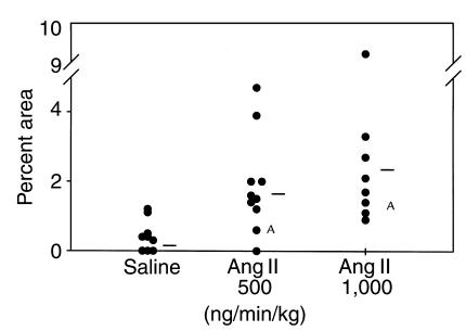
Figure 3.
Percent of intimal area covered by grossly discernible atherosclerotic lesions in the thoracic region. Circles represent the values for individual animals, and bars are the means for the groups. Infusion of Ang II significantly increased the percent of lesion area in the thoracic aorta. AStatistical difference of P > 0.05 from the control group using a Wilcoxon’s rank sum test.

