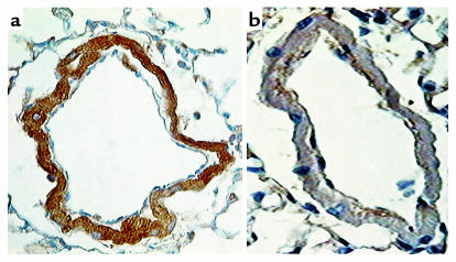
Figure 7.
Immunohistochemical localization of 5-HTT in distal pulmonary vessels from a 5-HTT+/+ mouse (a) and a 5-HTT–/– mutant mouse (b) exposed to hypoxia for 2 weeks. In the pulmonary vessels from wild-type mice, dense immunostaining is visible in the smooth muscle cells. In contrast, only a very low level of background staining is observed in vessels from 5-HTT–/– mice. Scale bar, 25 μm.

