Figure 4.
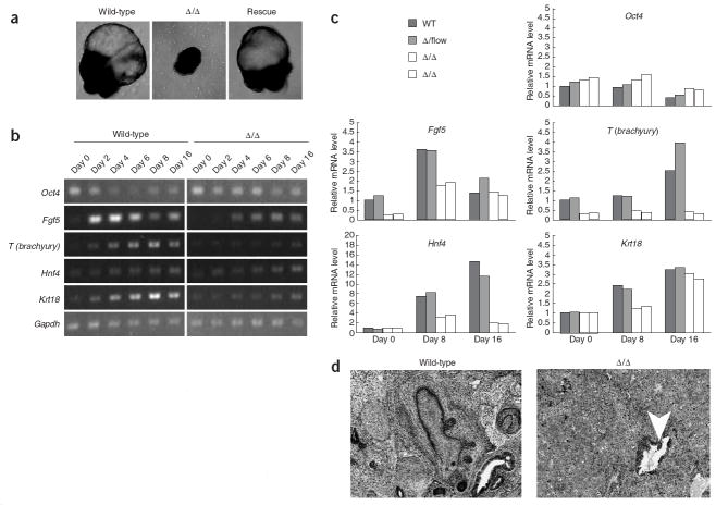
EB differentiation and teratoma formation. (a) Morphology of EBs from wild-type, Δ/Δ and rescued ES cells. EBs were cultured for 40–50 d. (b) RT-PCR analysis of pluripotency (Oct4) and differentiation markers (Fgf5,T(brachyury),Hnf4a,Krt18) after EB differentiation. Gapdh was used as a reference. (c) Quantitative RT-PCR for the markers shown in b. The β-actin gene was used as a reference. For each gene, data were normalized to the mRNA level at day 0 of wild-type EB differentiation. (d) Teratoma formation by wild-type and Dgcr8 knockout ES cells. Arrow in Δ/Δ tumor identifies region of epithelial differentiation.

