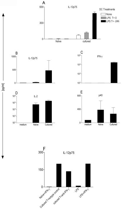
Figure 1. Antigen-activated but not naïve T cells induce IL-12p75 from DCs.
(A) BM-DCs at 2×10⁴ cells/well from B10.A-RAG2KO mice were cocultured for 48 hr with 1×10⁵ sorted naïve or antigen-activated 5C.C7 T cells +0.1μM MCC peptide in a 96-well U-bottom plate. LPS was added at 200ng/ml to DCs either at time zero (T=0) or 24h (T=−24h) and washed before the addition of T cells. ELISA (R&D system) specifically measured IL-12p75 in CSN after 48h. These data are expressed as the mean ± SD triplicates from a 96-well plate. (B–E) Multiplex cytokine array (SearchLight) measuring various cytokines in CSN obtained from LPS-activated (T=−24h) B10.A-RAG2KO. BMDCs at 2×10⁴ cells/well cocultured for 48 hr with 1×10⁵ naïve or antigen-activated 5C.C7 T cells +0.1μM MCC peptide in a 96 well U-bottom plate. These data are expressed as the mean ± SD composite of three independent experiments. (F) Same as (A) except two populations of DCs were used: 1-LPS-preactivated DCs (T=−24h) were either cocultured with naïve T cells plus 100ng/ml of rIFN-γ or with antigen-activated T cells plus 5μg/ml of neutralizing antibody for IFN-γ or 5μg/ml an isotype control Gt-anti-mouse. 2-“Resting” DC were stimulated with LPS or LPS plus IFN-γ stimulation.

