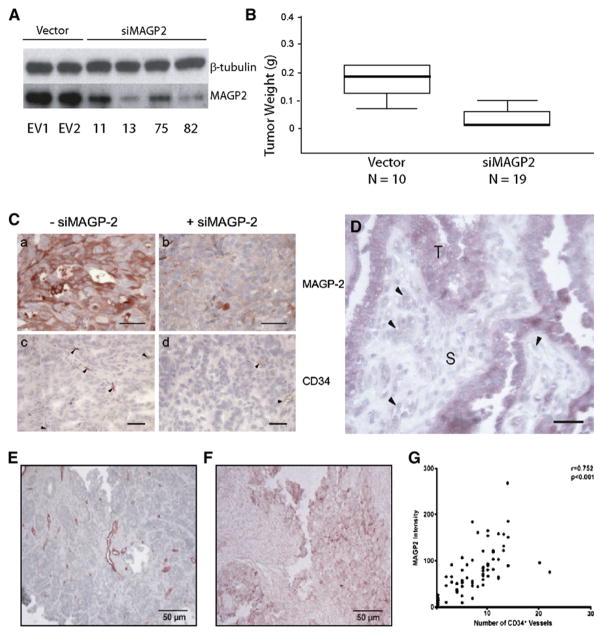
Figure 7. MAGP2 Expression Correlates with Tumor Size and CD34+ Microvessel Density in Serous Ovarian Cancer Tumors and Tissue Specimens.
(A) Western blot demonstrated knockdown of MAGP2 in five clones isolated from stably transfected SKOV3 cells, as well as three empty vector clones.
(B) MAGP2 knockdown resulted in significantly decreased tumor size in mice. The median weight of tumors from SKOV3 cells is 0.18 g; knockdown of MAGP2 results in a significant decrease in tumor weight (0.02 g; p < 0.01).
(C) MAGP2 knockdown resulted in decreased MAGP2 expression and CD34+ microvessel density in tumors developed in mice. Scale bars represent 50 μm.
(D) A section of a high-grade serous adenocarcinoma showing strong MAGP2 expression in tumor cells (T) but not in the stromal component (S) or the endothelial cells (arrowheads). The scale bar represents 100 μm.
(E) Immunolocalization of CD34+ microvessels in a human serous ovarian adenocarcinoma tumor section.
(F) MAGP2 expression in a late-stage high-grade human serous ovarian adenocarcinoma tumor section.
(G) Correlation between CD34+ and MAGP2 expression.

