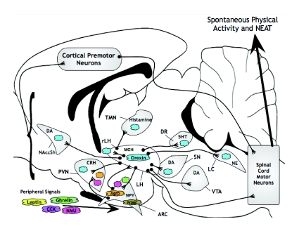
Fig. 5.
SPA (and resulting NEAT) regulatory brain areas and associated neuropeptides/transmitters [updated from fig. 1 in Kotz (Kotz, 2008)]. Colors correspond to specific neuropeptides/hormones as follows: blue, orexin; purple, CCK; pink, NMU; orange, Agrp; brown, POMC; green, ghrelin; yellow, leptin. Areas with these colors indicate site of synthesis (e.g. AgRP, POMC and ARC; orexin, LH), peripheral source (NMU, ghrelin, leptin and CCK), areas in which the neuropeptide/hormone has been injected and effects on SPA reported, or proposed site(s) of action (see text). Signals from all of these areas have the potential to influence cortical premotor neurons. Brain areas are not to scale, and connections and neuropeptides/transmitters indicated are not all-inclusive. Outline of rat brain was modified from Paxinos and Watson (Paxinos and Watson, 1990). For an alternative depiction, see fig. 2 in Castaneda et al. (Castaneda et al., 2005). 5HT, serotonin; Agrp, Agouti-related protein; ARC, hypothalamic arcuate nucleus; CCK, cholecystokinin; CRH, corticotrophin releasing hormone; DA, dopamine; LC, locus coeruleus; LH, lateral hypothalamus; MCH, melanin concentrating hormone; NAccSH, shell of nucleus accumbens; NE, norepinephrine; NMU, neuromedin U; NPY, neuropeptide Y; POMC, proopiomelanocortin; PVN, hypothalamic paraventricular nucleus; VTA, ventral tegmental area; rLH, rostral LH; SN, substantia nigra; TMN, tuberomammillary nucleus.

