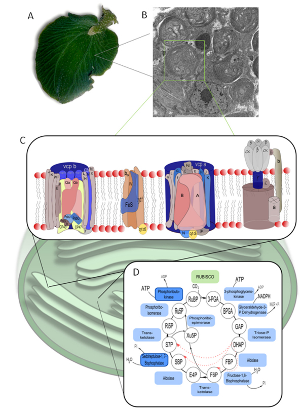
Fig. 5.
Schematic of the light and dark reactions of photosynthesis showing plastid- vs nuclear-encoded genes. (A) Adult, kleptoplastic Elysia chlorotica. (B) Transmission electron micrograph showing numerous algal plastids within a cell lining the digestive diverticuli of the sea slug. (C,D) Schematic of the two photosynthetic processes overlaid on a plastid illustrating the essential proteins required in each pathway. Nuclear-encoded plastid proteins are shaded blue for both the electron transfer chain (C) and the Calvin–Bensen cycle (D). In the latter, RuBisCO is shaded green to indicate a plastid-encoded protein. Two of the enzymes, phosphoribulokinase and sedoheptulose-1,7-bisphosphatase, are shaded dark blue to indicate that, although they are nuclear-encoded like the light-blue-shaded enzymes, these enzymes are unique to phototrophs and are not typically found in an animal, whereas the light-blue-shaded enzymes all have homologs in animal metabolism.

