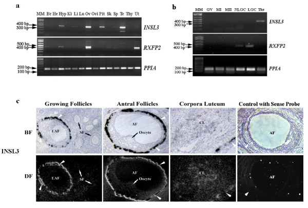
Figure 2.
Detection of INSL3 and RXFP2 transcripts in rhesus macaques. a. Tissue distribution of INSL3-RXFP2 in the rhesus macaque. Total RNA extracted from the brain cortex (Br), heart (He), hypothalamus (Hyp), kidney (Ki), liver (Li), lung (Lu), ovary (Ov), oviduct (Ovi), pituitary (Pit), skeleton muscle (Sk), spleen (Sp), testis (Te), thymus (Thy) and uterus (Ut) were subjected to RT-PCR. b. Cellular expression of INSL3-RXFP2 in the macaque ovary. GV: germinal vesicle-intact oocytes; MI, MII: metaphase I, II oocytes; NLGC, LGC: non-luteinized, luteinized granulosa cells; The: Theca cells. PPIA was used as an internal control and 1 kb plus DNA ladder (Invitrogen) was used as molecular marker (MM) in both cases. c. In situ hybridization of INSL3 in the monkey ovary. White arrowheads denote theca layers of antral follicles. SF, secondary follicle; EAF: early antral follicle; AF: antral follicle; CL: corpora luteum; BF: bright field; DF: dark field. All experimental samples were collected from at least 3 individual animals.

