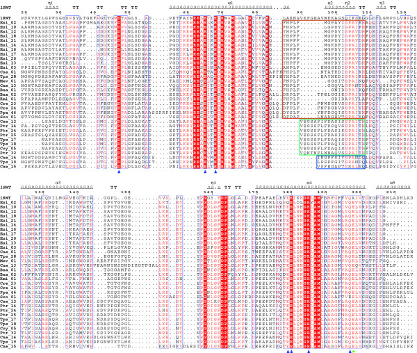
Figure 3.
Structure-based sequence alignment of the crystallized spinach CAB (code 1RWT) with proteins belonging to the LI818 clade. The secondary structure of the spinach CAB is shown above the alignment. Conserved amino acids highlighted by a red background are identical and those in red letters are similar. Alpha helices are represented as helices, and β-turns are marked with TT. Blue triangles indicate the conserved residues involved in the binding of chlorophyll a molecules. The green star shows the conserved glutamate in LI818-like proteins, predicted to preclude the binding of Chlb 607 observed in the spinach CAB. The colored frames indicate the three subgroups of helix α2 within the LI818 subfamilies.

