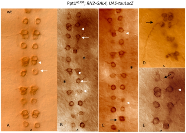
Figure 3. Ppt1 mutants exhibit abnormal RP2 axon trajectories.
S16 ventral nerve cord stained with anti-β-gal to detect RN2-tau-lacZ expressed in aCC, pCC and RP2 neurons. (A) Wild type RN2-tau-lacZ pattern showing RP2 neuron and its axon trajectory (white arrow); and aCC/pCC neurons (arrowhead). (B–D). Ppt1A179T; RN2-Gal4:UAS-tauLacZ embryos display a loss of RP2 motoneurons (asterisks) and aCC/pCC neurons (arrowheads) in many hemisegments; and disorganized cellular arrangements of the remaining LacZ-positive neurons. Many hemisegments have normal RP2 axon trajectory (white arrow) while others do not (black arrow). The RP2 in panel C shows an abnormal axon trajectory projecting posteriorly instead of anteriorly. Panel E is an enlarged subset of B. Anterior, up; caret, midline.

