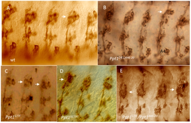
Figure 6. Chordotonal neurons (lch5) in the developing PNS are abnormal in many Ppt1 LOF embryos.
In all panels, lch5 neurons are indicated by the arrows. Side view of S17 embryos stained with Mab 22C10. (A) Wild type clusters of lch5, v, and v′ neurons are located in every abdominal segment. (B–E) lch5 neurons in mutant Ppt1 embryos demonstrate a variety of defects: decreased number of neurons (B), fused and abnormally shaped (D, E), organization and positional defects, and thin axon bundles (black arrowhead) (B and C). dorsal, up.

