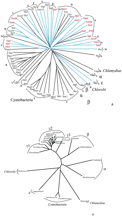
Figure 1. Phylogenetic tree of the full-length Int/Inv proteins.
LysM containing proteins are colored in red, and proteins possessing paired cysteines, with the capacity to form disulphide bonds, are indicated with blue branches. Clusters A to X were analyzed for sequence conservation (see text), as indicated in the figures. This tree, and those presented in figures 2 and 3 are based on CLUSTAL-X-derived multiple alignments shown in Figures S1, S2 and S3, respectively. The trees were drawn with the TreeView program [34]. The organismal origins of the proteins are indicated adjacent to the branch/cluster number except for the large majority of proteins from the γ-proteobacteria which are unlabeled. This convention is also used in figures 2 and 3. Using the same program, the tree for the ribosomal RNAs, corresponding to the represented genera, was derived for the second part of this figure.

