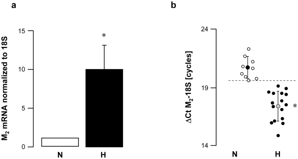
Figure 2. M2 muscarinic receptor gene expression in peripheral mononuclear white blood cells from normal (N) and vagal hyperreactive (H) rabbits.
R-R intervals were measured in conscious rabbits challenged with PNE 500 µg kg−1 following the procedure described in Material and Methods. M2 muscarinic gene expression was assessed in peripheral mononuclear white blood cells by quantitative RT-PCR and normalized to the rabbit 18S housekeeping gene. Values in (a) show amplification ratio calculated according to the 2−ΔΔCt method of 9 (N) and 16 (H) experiments. In (b), each symbol represents one animal; ΔCt M2-18S corresponds to the number of amplification cycles needed to detect M2 fluorescence standardized to 18S; thus, the lower the ΔCt M2-18S, the greater M2 mRNA initial quantity. *: P<0.0001 versus N.

