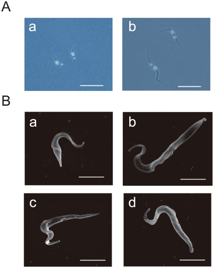
Figure 6. The morphology of TbUNC119-TbUNC119BP double knock-down cells.
A. Fluorescent differential interference contrast image of TbUNC119-TbUNC119BP double knock-down procyclic form cells. TbUNC119-TbUNC119BP double knock-down cells 7 days after induction (b) and RNAi-uninduced control cells (a) were labeled with DAPI. Note that the posterior end and the kinetoplast were extended in the RNAi-induced double knock-down cells. The phenotype was clearly observed from 5 days after RNAi induction in double knock-down cells. Bar = 20 µm. B. Scanning electron microscopy of TbUNC119-TbUNC119BP double knock-down procyclic form cells. Double knock-down cells 7 days after RNAi induction (b–d) and RNAi-uninduced control cell (a) were used for the analysis. Bar = 10 µm.

