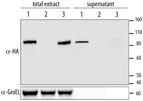
Figure 2. Expression analysis of XopD.
Strains 85* (XopD-HA) (1), 85* (2) and 85* ΔhrcV (XopD-HA) (3) were incubated in MOKA rich medium (total extract, left) or secretion medium (supernatant, right). Total protein extracts (10-fold concentrated) and TCA-precipitated filtered supernatants (200-fold concentrated) were analyzed by immunoblotting using anti-HA antibodies (upper panel) to detect the presence of XopD, or anti-GroEL antibodies (lower panel) to show that bacterial lysis had not occurred.

