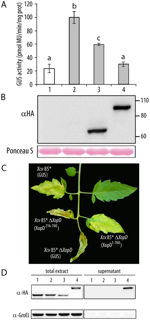
Figure 6. In planta analysis of XopD1-760-mediated virulence functions.
(A) Transactivation of the PR1 promoter after SA treatment in transient assays in N. benthamiana. Leaves were inoculated with A. tumefaciens carrying a 35S:PR1p-GUS fusion either alone (lanes 1, 2) or together with HA-tagged XopD216-760 (lane 3) or XopD1-760 (lane 4). 18 hours after agroinfiltration, leaves were mock-treated (white bar) or treated with 2 mM SA (grey bars). Fluorimetric GUS assays in leaf discs were performed 12 hours later. Mean values and SEM values were calculated from the results of four independent experiments, with four replicates per experiment. Statistical differences according to a Student's t test P value <0.05 are indicated by letters. MU, methylumbelliferone. (B) Western blot analysis showing expression of HA-tagged XopD216-760 and XopD1-760. Ponceau S staining illustrates equal loading. (C) Susceptible Pearson tomato leaves were inoculated with Xcv 85* or Xcv 85* ΔxopD, expressing an HA-tagged GUS control, Xcv 85* ΔxopD expressing HA-tagged XopD216-760 or Xcv 85* ΔxopD expressing HA-tagged XopD1-760. Inoculation was performed with bacterial suspensions of 1×10⁵ cfu/ml. Representative symptoms observed 10 dpi are shown. Similar phenotypes were observed in four independent experiments. (D) Strains Xcv 85* expressing a GUS control (1) and 85* ΔxopD expressing either a GUS control (2), XopD216-760 (3) or XopD1-760 (4) were incubated in MOKA rich medium (total extract, left) or secretion medium (supernatant, right). Total protein extracts (10-fold concentrated) and TCA-precipitated filtered supernatants concentrated (200-fold concentrated) were analysed by immunoblotting using anti-HA antibodies (upper panel) to detect the presence of GUS, XopD216-760 and XopD1-760, or anti-GroEL antibodies (lower panel) to show that bacterial lysis had not occurred.

