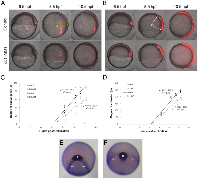
Figure 8. Knockdown of cfl1 causes convergent and extension defects.
(A, B) Embryos were injected with Q-rhodamine with or without cfl1 tMO1 and incubated in the dark. Cells of the lateral (A) and dorsal blastomere margins (B) are marked as shown in red fluorescence at the shield stage and observed until 10.5 hpf. Representative photographs taken at 6.5, 8.5 and 10.5 hpf for both the untreated control and cfl1 MO-injected embryos are shown. (C) As shown in the panel A with an 8.5-hpf control embryo, the radius of the embryo (R) and the migration distance of the labeled cells (r) were measured. The degree of convergence (θ) was calculated by applying the following equation: θ = sin−1(r/R), and then plotted against the stage of the embryo in hpf. (D) As shown in panel B with an 8.5-hpf control embryo, the angle (φ) between the two arrows of the anterior and posterior ends of the marked axial mesoendoderm connecting the center of the embryo was measured in each embryo to estimate the degree of extension. The degree of extension (φ) was then plotted against the stage of the embryo. (E, F) WISH against cstl1b (expressed in the prechordal plate as indicated by asterisks) and dlx3b (expressed in paraxial mesoderm as indicated by arrows) of bud-stage zebrafish embryos was performed. Representative photographs in frontal view are shown for an untreated (E) and a cfl1 tMO1-injected embryo (F).

