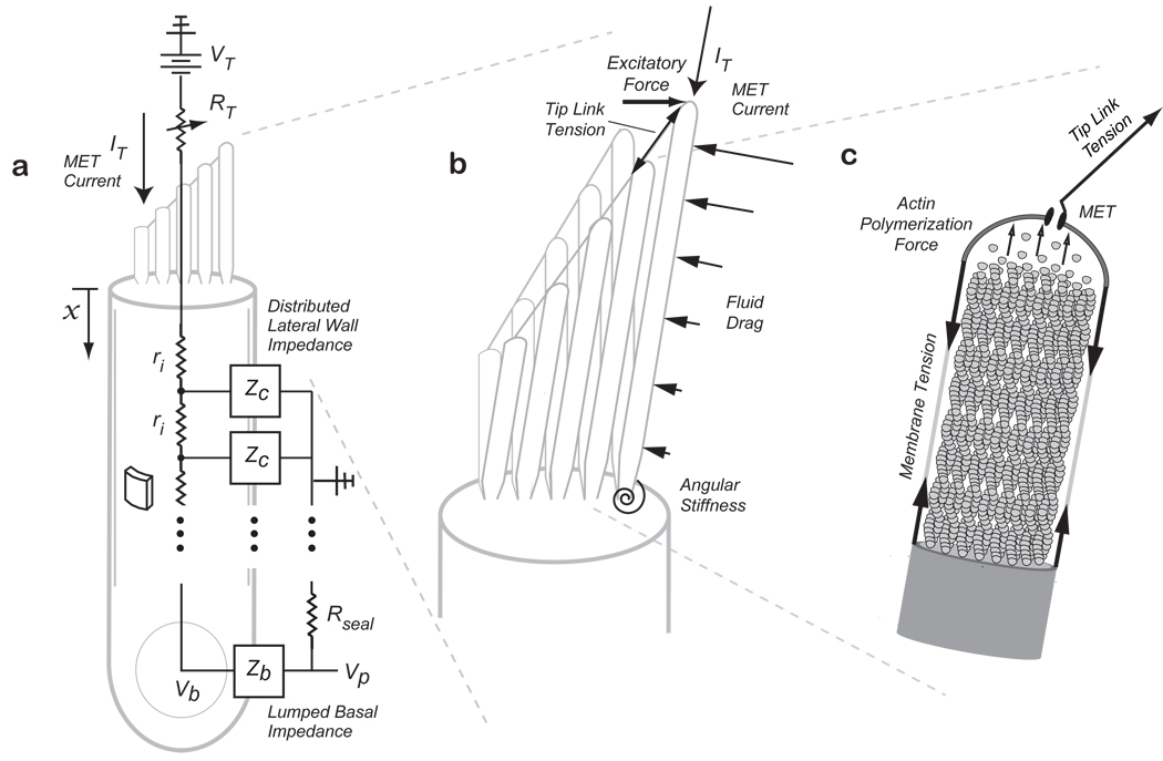
Figure 2.
Model Schematic showing (a) somatic piezoelectricity represented as a distributed lateral wall piezoelectric element ZC, with an intracellular fluid resistance, ri, and a lumped basal impedance, Zb. Input MET current, IT, enters the apically positioned stereocilia bundle with a variable resistance at their tips representing the MET. Stereocilia bundle flexoelectricity (b) is represented as stereocilia connected by tip-links with input MET current at their tips. An excitatory force tilts the bundle and is resisted by tension in the tip-links, fluid-drag forces along the length of the stereocilia, and angular stiffness at the base. The stereocilia core is filled with actin filaments (c) and the actin polymerization force required to maintain the stereocilium height generates the resting membrane tension (based on 14,16).

