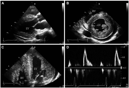
Fig. 3.
Two-dimensional echocardiography reveals severe concentric LV hypertrophy, mild LV systolic dysfunction, a small pericardial effusion, and increased RV free wall thickness with mild RV systolic dysfunction. A: parasternal long axis view. B: parasternal short axis view at the mid-ventricular level. C: magnified apical 4-chamber view. D: mitral inflow view (E velocity, 90.28 cm/s; A velocity, 36.77 cm/s; deceleration time, 216.64 ms; E/E': 33.4). LV: left ventricular, RV: right ventricular.

