
Figure 4. Native ScTx1-sensitive Kv current of RMCA myocytes.
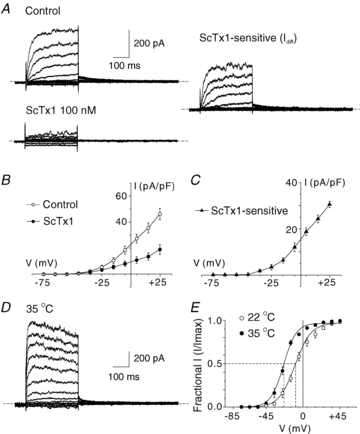
A, representative recordings of whole-cell Kv current of an RMCA myocyte in the absence (Control) and presence of 100 nm ScTx1 (left) and the ScTx1-sensitive current (right) determined by digital subtraction of ScTx1 from Control current at 22°C. Voltage steps of 325 ms duration between −95 and +45 mV in increments of 10 mV prior to repolarization to −45 mV were applied from a holding potential of −75 mV. Similar recordings were obtained from 7 additional myocytes from cell isolations of RMCAs of 3 rats. B and C, mean values ± s.e.m. (n = 6) for net whole-cell and ScTx1-sensitive Kv current I–V relations. D, representative recording of RMCA ScTx1-sensitive Kv current at 35°C. E, mean values ± s.e.m. for normalized tail current amplitude versus command step voltage for native current at 22 and 35°C (n = 6 and 3, respectively) that exhibited complete suppression of tail currents following treatment with ScTx1. Continuous lines represent best fits to the data points using a standard Boltzmann function.