
Figure 8. PLA detection of plasma membrane expression of recombinant Kv channel protein in HEK 293 cells.
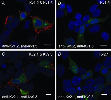
A, PLA reaction product indicated by red fluorescent dots was detected at the periphery of GFP-positive HEK 293 cells transfected with cDNAs encoding GFP, Kv1.2 and Kv1.5, but not in non-transfected GFP-negative cells probed with Kv1.2 and Kv1.5 primary antibodies. Here and in subsequent panels, the nuclei of GFP-positive and -negative cells are indicated by the blue Hoechst 33342 stain, the Kv channel cDNAs and primary antibodies used are indicated in the upper right and lower left corners, respectively, the scale bars are 10 μm in length and each image is an optical section of 0.3–0.5 μm thickness at a mid-cell depth. B, lack of PLA reaction product at cell periphery of GFP-positive cells transfected with Kv1.5 only (i.e. no Kv1.2) and probed with Kv1.2 and Kv1.5 primary antibodies. C, PLA signals were detected at the periphery of GFP-positive cells transfected with Kv2.1 and Kv9.3, but not in GFP-negative cells probed with Kv2.1 and Kv9.3 primary antibodies. D, lack of PLA signals at cell periphery of GFP-positive cells transfected with Kv2.1 (i.e. no Kv9.3) and probed with Kv2.1 and Kv9.3 primary antibodies.