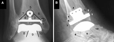
Fig. 2A–B.
(A) AP and (B) lateral postoperative radiographs show the different zones of cysts for the tibial and talar components. On the AP view, the tibia is divided into seven zones (1 = medial malleolus, 2 = medial flange, 3 = medial tibial base plate, 4 = lateral tibial base plate, 5 = tibial keel, 6 = medial tibial metaphysis, and 7 = lateral tibial metaphysis), whereas the talus consists of three zones (8 = from the lateral talar flange to the lateral aspect of the talar keel, 9 = directly under the talar keel, and 10 = from the medial aspect of the talar keel to the most medial aspect of the talar component). On the lateral view, the tibia is divided into seven zones (1 = anterior tibia, 2 = anterior tibial base plate, 3 = anterior keel stem, 4 = tibial keel, 5 = posterior keel stem, 6 = posterior tibial base plate, and 7 = posterior tibia), whereas the talus consists of three zones (8 = from the posterior extreme of the talar component to the posterior aspect of the talar keel, 9 = under the talar keel, and 10 = from the anterior aspect of the talar keel to anterior extreme of the talar component).

