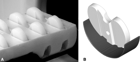
Fig. 1A–B.
(A) Photograph of inserts held in foam on the bed of the scanner and (B) a rendered image of the insert in the bed at a double-oblique angle of approximately 10° backward vertically and 3° laterally. The foam that held the inserts is radiotranslucent and does not appear in the reconstructed scan images.

