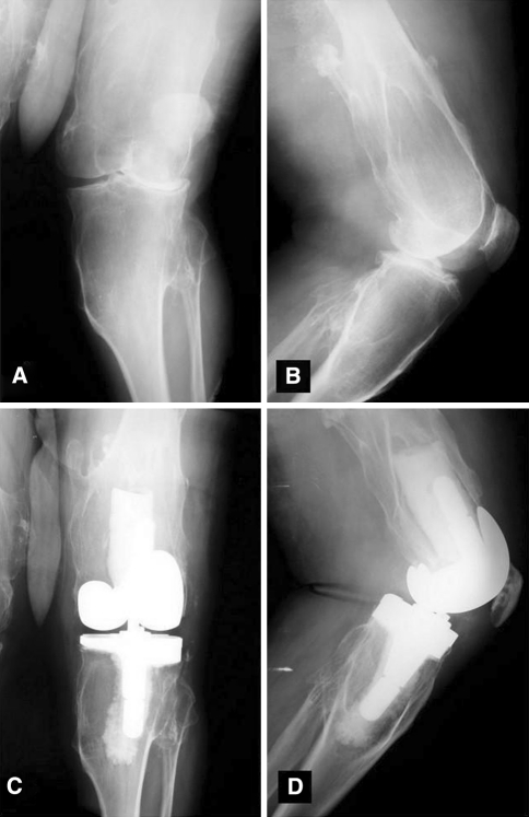
Fig. 1A–D.
Preoperative (A) AP and (B) lateral radiographs of a patient with multiple hereditary exostosis demonstrate exostoses and valgus alignment. Postoperative (C) AP and (D) lateral radiographs demonstrate corrected deformity with use of a varus-valgus constraining implant and lateral tibial augment.

