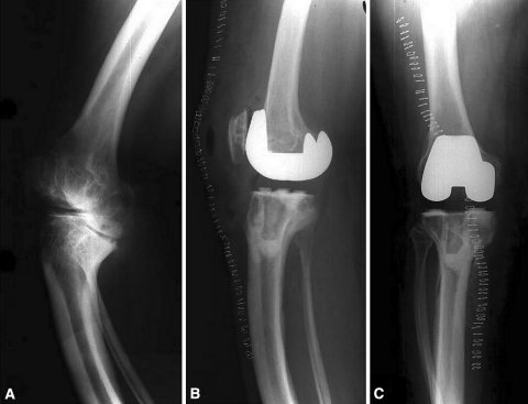
Fig. 3A–C.
(A) A preoperative radiograph (oblique view as a result of torsional deformity) of a patient with osteogenesis imperfecta demonstrates contorted tibial geometry. Postoperative (B) AP and (C) lateral radiographs demonstrate use of an all-polyethylene tibial component with modification of the tibial stem to accommodate the tibial deformity.

