
Figure 5b:
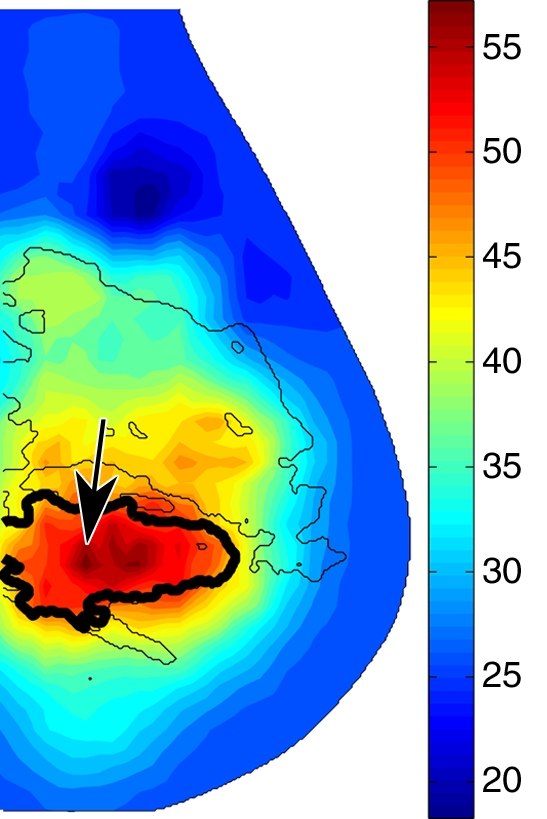
Reconstructed image sections for the right breast in 45-year-old woman. (a) DBT image and (b) HbT (micromoles per liter), (c) So2, and (d) μs′ (cm−1) images at 830 nm. The breast contains a 2.5-cm invasive ductal carcinoma (arrow on a, line with arrow on b-d).