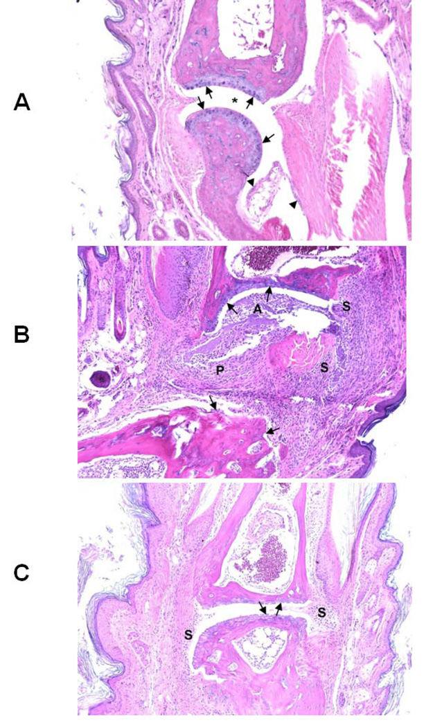
Figure 10.
Representative tissue sections taken from G1, G2 and G4 group of mice (see Table 2 for grouping) are shown in the figure. A) Normal phalangeal joints of hind foot of control mouse without intradermal injection of type II collagen (CII) (4 mice in each group). Cartilaginous surfaces are intact and smooth (arrows) with normal synovium (arrowheads) and clear joint space (*). B) Severely arthritic phalangeal joint of hind foot of mouse intradermally injected with CII without further treatment. Marked cartilaginous and bone erosion (arrows) are present with severe pannus formation (P) and synovitis (S). The articular space (A) is filled with tissue detritus, fibrin, and mixed inflammatory cells. C) Mildly arthritic phalangeal joint of hind foot of mouse intradermally injected with CII and treated with P3. Both cartilaginous surfaces of articulating phalanges are eroded (arrows) with slight synovial hyperplasia (S) but lacking both invasive pannus and significant inflammatory exudate. H&E staining. Original magnification ×100.

