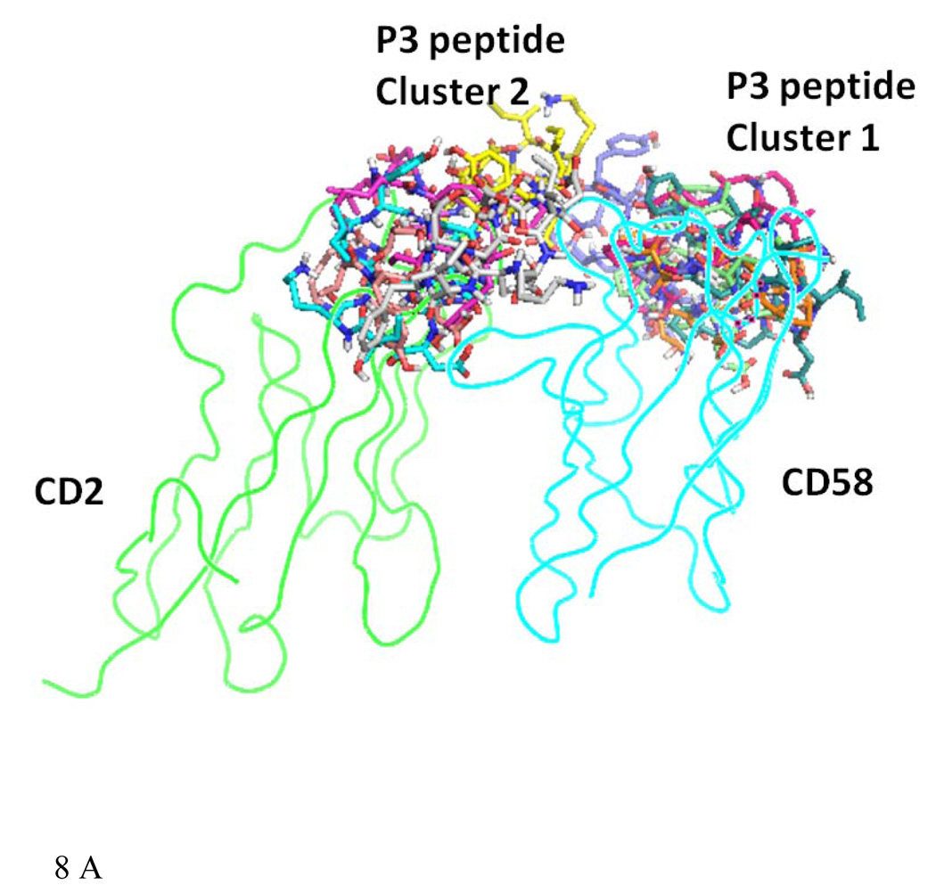
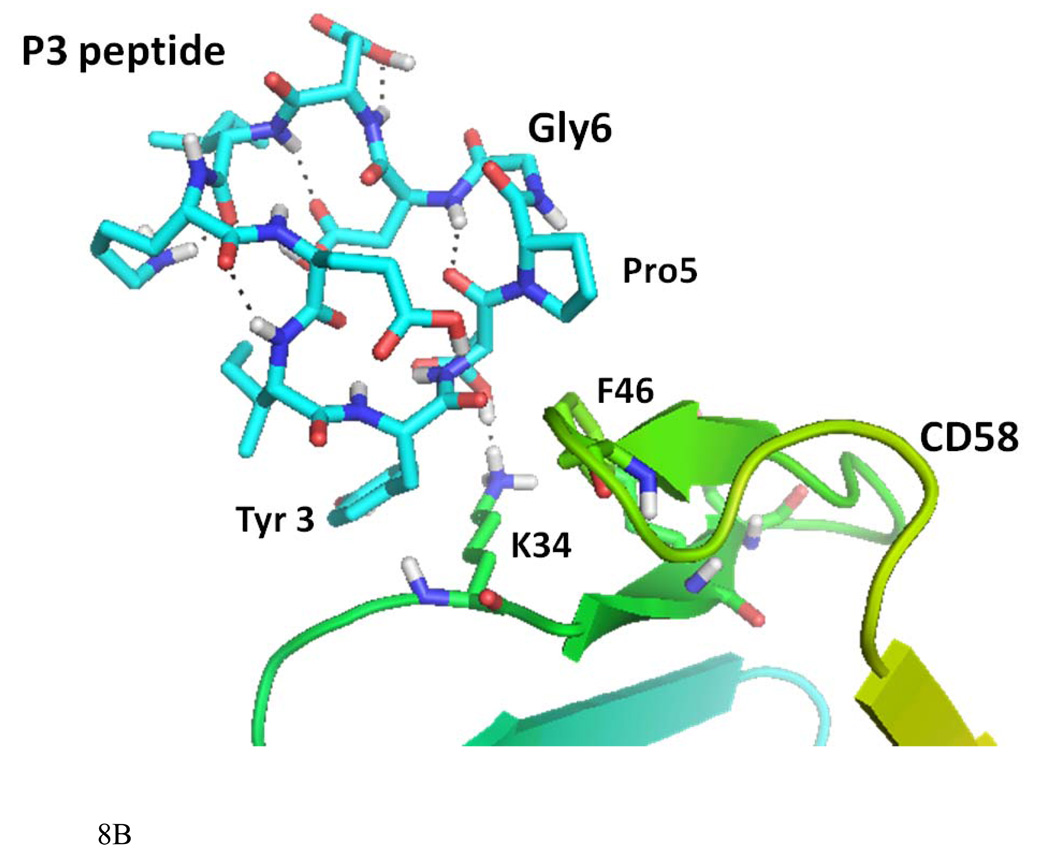
Figure 8.
A) Docked structure of P3 to CD58 protein domain D1. Two clusters with low docking energy are shown in the figure. B) Docked structure of P3 on the adhesion domain of CD58. Detailed interactions of P3 and CD58 protein are shown here. Amino acids of the peptide are labeled with three letter code, and amino acids of protein CD58 are labeled with single letter code for clarity. Interaction of Tyr3 and Asp4 and Pro5 of the peptide with K34 and F46 of protein CD58 are shown.


