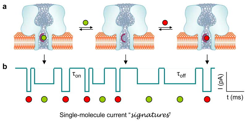
Figure 1.
Single-molecule detection in a protein pore. (a) Competitive and reversible binding of different analytes (represented by green and red balls) to a receptor engineered in the protein pore. (b) Stochastic current blocks function as “signatures” of single bound analytes, which, based on block amplitude and duration, allow for the identification of the unknown analytes. In the diagram, the red analyte blocks more current with a shorter duration than the green one. Analytes can also be quantified by their block occurrence.

