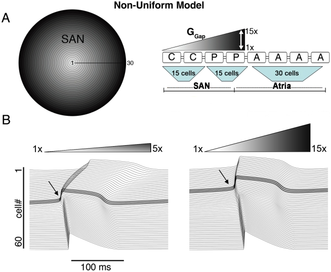
Figure 1. The non-uniform model.
A) The non-uniform model schematic and its equivalent 1D strand with various levels of gradients in coupling is shown (‘C’ central cell, ‘P’ peripheral cell, ‘A’ atrial cell and ‘GGap’ intercellular coupling). B) The site of pacemaker initiation (bold and arrow) under conditions of shallow (left) and steep (right) GGap gradient (respectively).

