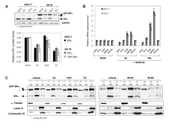
Figure 2.
Nuclear accumulation and degradation of the estrogen receptor alpha in MCF7 and SK19 cells. A) Protein levels of endogenous ERα and GFP-ERα in MCF-7 and SK19 cells in response to E2 and to anti-estrogens. The SK19 cell line was generated from MCF-7 cells by stably transfecting a GFP-ERα expression vector (pEGFP-C2-hERα). B) Total RNA was extracted from SK19 and MCF-7 cells treated or not for 2 h and 16 h with 10 nM E2. Relative expression level of the ESR1, TFF1, GREB1 and PGR genes in SK19 cells was analyzed by qRT-PCR and compared to gene-expression regulation in MCF-7 cells. RPLP0 served as an internal control (see Methods). Data shown are an average of two independent experiments, error bars represent ± S.E. mean. C) Cellular distribution of ERα and GFP-ERα from digitonin based cellular fractionation experiments. SK19 cells were treated with drugs for 3 h. Nuclear fractions of untreated and treated cells with 10 nM E2, 1 μM OHT, 1 μM ICI, 1 μM RU39 and 1 μM RU58, were isolated as described in "Methods". Nuclear content of ERα and GFP-ERα was analyzed by Western Blotting using anti-ERα antibodies. The specific subcellular proteins, α-tubulin for cytoplasmic fraction (Cyt), Lamin A for nuclear insoluble fraction (NI) and cytokeratin 18 for nuclear soluble fraction (NS) are loading controls for the different cellular compartments. Results shown are representative of at least 3 independent experiments.

