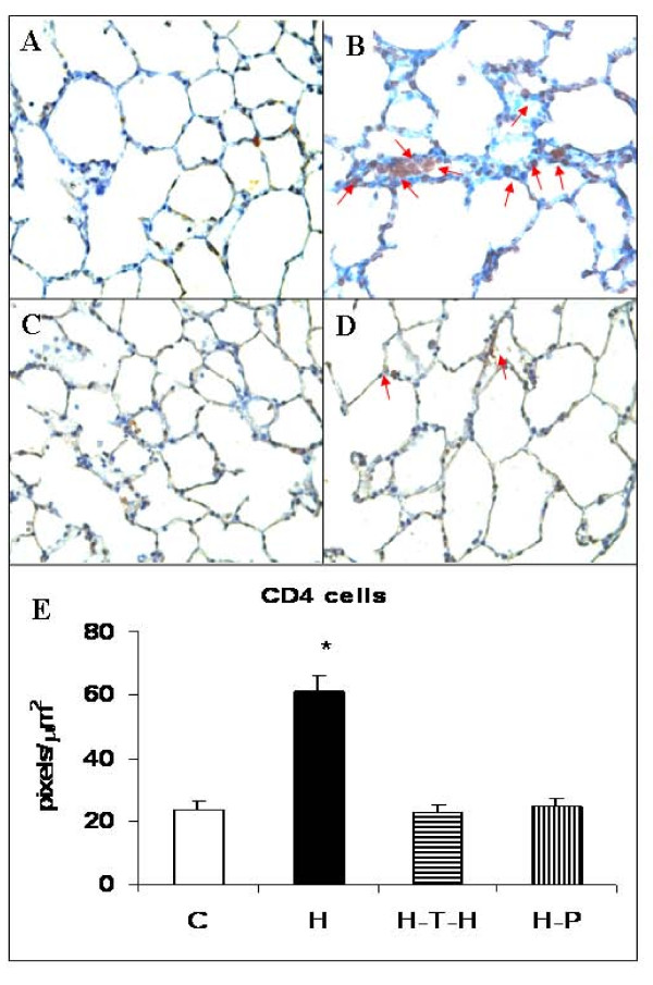
Figure 4.
Immunostaining for CD4+ T cells. Immunohistochemistry for CD4+ T cells in PBS-treated (A); HUVEC-immunized (B); intrathymically HUVEC-injected and HUVEC-immunized (C); and pristane-treated 3 days prior to HUVEC-immunization (D) rat lung sections. Magnification x400. (E) The CD4+ cell accumulation areas were measured in pixels per square micrometers. A macro was created to automate the segmentation, processing, and quantification steps. For each animal (n = 4), 10 fields at a magnification of x100 were captured in a blinded fashion using Carl Zeiss KS300 Imaging System software. *p ≤ 0.002 when compared to immune tolerized rats.

