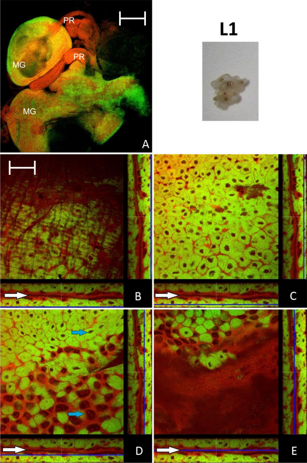
Figure 1.
Larva of stage L1. A: Overview showing two midguts (MG) and their proventriculi (PR) by confocal laser scanning microscopy. B - E: Four orthogonal views of confocal image stacks of C floridanus L1 larva midgut sections. The blue lines in the XZ and YZ stack representations (below and on the right side of each quadratic micrograph) illustrate the position of the image plane (XY). The bacteria-free midgut cells typically have large nuclei and several nucleoli while the bacteriocytes are characterized by small nuclei (blue arrows in D). The bacteriocytes form a nearly contiguous layer surrounding the midgut (B, C) directly underneath of the muscle network (A and Fig. 3). There are no bacteriocytes present in the cell layer lining the midgut lumen (D, E). The midgut lumen is indicated by white arrows. Green label: The Blochmannia specific probe Bfl172-FITC; red label: SYTO Orange 83. The scale bars correspond to 220 μM (A) and 35 μM (B - E), respectively.

