Figure 1.
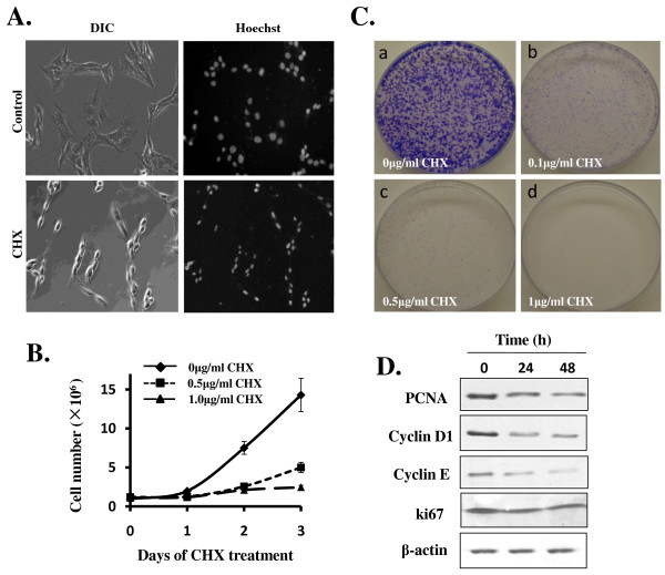
LCC potently inhibits C6 glioma cell proliferation. A) C6 cells were seeded in 6-well plates and treated without or with CHX (1 μg/ml) for 48 h. For observation of the integrity of nuclei, cells were stained with Hoechst 33258 and visualized under fluorescence microscope. Both phase contrast (left panels) and fluorescent (right panels) photos were shown (magnification: ×200). B) C6 cells were seeded in 6-well plates at a density of 1.0×10⁵ per well. CHX were added at concentration of 0 (◆) (as control), 0.5 μg/ml(■), and 1 μg/ml(▲), as indicated. Cells were collected in each of the next 3 days after trypsin treatment and cell numbers were counted using a haemocytometer. Data are presented as means ± SEM (n = 3). C) Colony Formation Efficiency Assay. 1,000 cells were plated in 35-mm tissue culture dishes. After overnight culturing, cells were exposed to different doses of CHX: (a) control; (b) CHX 0.1 μg/ml; (c) CHX 0.5 μg/ml; (d) CHX 1 μg/ml. Photos were taken 6 days later after staining with 0.5% crystal violet. d) Western immunoblotting analysis on cell cycle regulators and indicators. C6 Cells were cultured in 6-well plates and were treated without or with CHX (0.5 μg/ml) for indicated time. Western blotting was performed as described in the Methods.

