Figure 3.
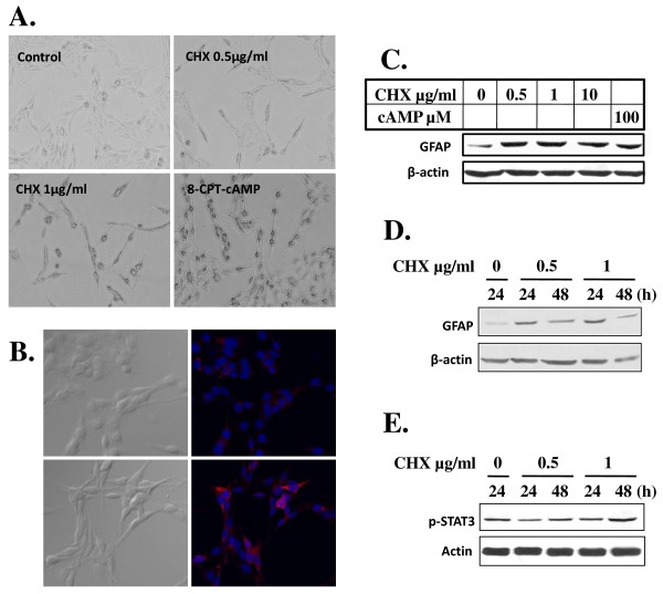
LCC induces C6 cell differentiation. A) LCC promotes morphological transformation of C6 cells that indicated cell differentiation. C6 cells were exposed to CHX at indicated concentration and to 8-cpt-cAMP (100 μM) for 24 h. Photos were taken as in Figure 1A (magnification, ×200). B-D) Treatment of C6 cells with LCC upregulates GFAP. In B), C6 cells cultured for 12 h in the absence (upper panels) or presence (lower panels) of CHX (1 μg/ml) were either photographed directly (left panels) or immunostained for GFAP (red) as described in Materials and Methods. Nuclei were stained with Hoechst 33258 as in Figure 1A. In C and D), GFAP expression was assessed in Western immunostaining analysis. Cells were treated with CHX at indicated concentration for 24 h (C) or 24 and 48 h (D). β-actin was used as loading control. E) Western blotting was done as in D) except anti-p-STAT3 was used. β-actin was used as loading control.

