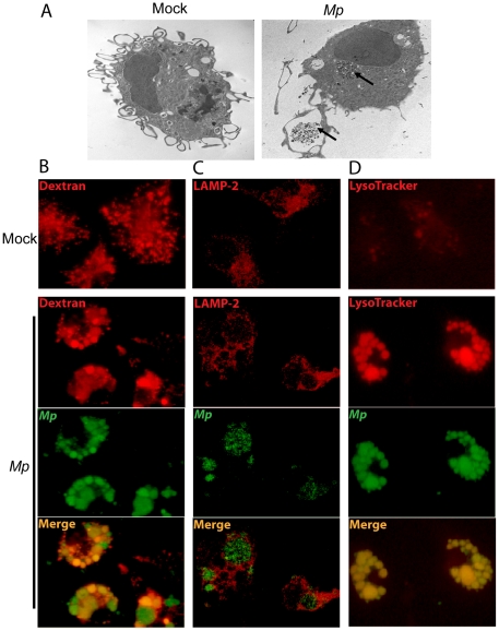
Figure 5. Macrophage phagocytosis of Mp in vitro.
(A) BMM were infected with Mp for 1 h, and the cells were fixed with 2% paraformaldehyde and 2% glutaraldehyde prior to embedding for TEM. Internalized Mp are highlighted by the black arrows. BMM were pre-loaded with dextran to label endosomes (B), and stained with anti-LAMP-2 antibody to visualize lysosomes (C) or LysoTracker dye to visualize acidified compartments (D). (B-D) cultures were infected with Mp (MOI 100∶1) or mock infected with medium alone as indicated. All portions of these experiments were repeated at least two times, always with similar results.

