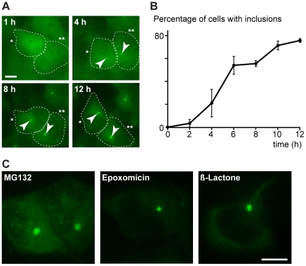
Figure 1. Ubiquitin-containing aggregates caused by proteasome inhibition.
(A) Formation of aggregates (arrowheads) induced with 20 µM MG132 was monitored by live-imaging a SH-SY5Y cell population that stably expresses GFP-ubiquitin. Two representative cells (indicated with one and two asterisks) are shown at different times. Scale bar represents 10 µm. (B) Percentage of methanol-fixed GFP-ubiquitin SH-SY5Y cells containing at least one GFP-ubiquitin inclusion (defined by a four fold increase of GFP signal intensity) after the addition of 20 µM MG132 for the indicated times. Average of three experiments with standard errors are shown (n = 200). (C) Representative methanol-fixed GFP-ubiquitin cells treated with 10 µM MG132, 1 µM epoxomycin or 2 µM clasto-lactacystin β-lactone for 12 h. Scale bar represents 10 µm.

