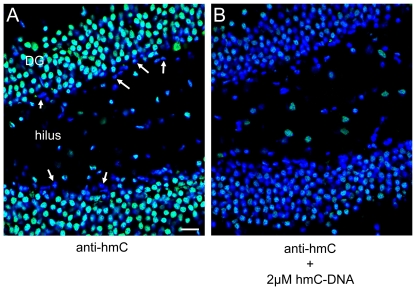
Figure 6. Immunolocalization of hmC in mouse hippocampus.
High magnification images of hmC immunoreactivity in the dentate gyrus (DG) and the hilus of mouse hippocampus. A) Signal for anti-hmC (green) and Hoechst 33342 nuclear dye (blue) B) Competition of anti-hmC with 2 µM hmC-DNA. The scale bar marks 20 µm.

