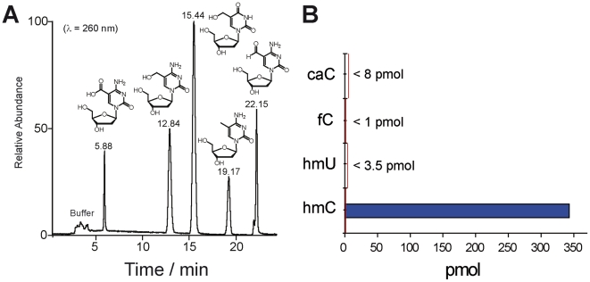
Figure 7. Detection of potential demethylation intermediates caC, hmU, and fC.
A) HPLC-chromatogram of the synthesized cytosine and uracil modifications caC, hmC, hmU, mC, and fC as 2′-deoxynucleosides showing excellent separation of the compounds. B) Detected values of the potential intermediates as example in olfactory bulb. The red line indicates the detection limits of the modified nucleosides in enzymatically digested DNA samples.

