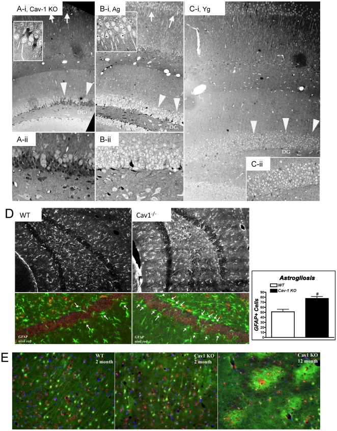
Figure 5. Cav-1 KO mice exhibit enhanced astrogliosis and neuronal degeneration.
(A–C) Light microscopic image displaying 0.5 µm thick hippocampal sections of Cav-1 KO (A -i, A -ii), aged (B -i, B -ii), and young (C -i, C -ii) stained with toludine blue. There is a drastic reduction in neurons within the dentate gyrus (large arrow heads) and CA1 regions (arrows) of young Cav-1 KO mice compared to young and aged WT. In addition, there appears to be the presence of more glia and glial scar formation within the dentate gyrus of Cav-1 KO mice as indicated by the darker gray cell bodies intermixed with the neurons. (D) Hippocampal coronal cryostat sections (10 µm) from WT and Cav-1 KO mice were stained with Nissl (neuronal marker, red pixels) and GFAP (astrocyte marker, green) to show no overlap between neurons and astrocytes. (E) Coronal cryostat sections (25 µm) of 2 month WT, 2 month Cav-1 KO and 12 month Cav-1 KO stained with 0.0004% Flouro-Jade®B and fluorescent red Nissl with DAPI. Areas from CA1 of the hippocampus were imaged. WT CA1 showed well-organized astrocytes. Two month Cav-1 KO had areas of disorganized astrocytes with lightly labeling areas of potential future plaque development. Twelve month Cav-1 KO CA1 areas had large bright, entangled green fluorescence with red neurons inside and significantly less organized astrocytes, further demonstrating a degenerating neuronal model.

