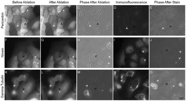
Figure 1. Immunofluorescence staining with percentrin, ninein, and gamma tubulin of centrosome irradiated cells.
The laser was exposed to the centrosome region as identified by the presence GFP fluorescence in A,F,K. Following laser exposure a successful irradiation event was determined by loss of fluorescence or fragmentation of the centrosomal marker signal at the position of irradiation (black arrow). Three different centrosomal probes were used where 27 of 28 pericentrin stained cells, 22 of 24 ninein stained cells, and 19 of 20 gamma tubulin stained cells were determined successful. Both GFP and immunofluorescence signal of centrosomes in neighboring control cells were not affected following laser exposure as pointed out by the white arrows in B, G, and L. Phase images after irradiation (C,H, and M) were matched following the staining procedure (E, J, and O).

